A stinkbug (Megymenum gracilicorne) mother covers her eggs with fungal hyphae
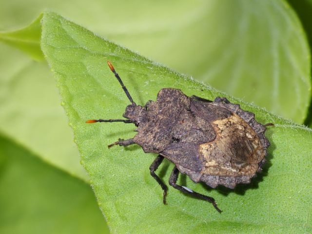
Females of the stinkbug Megymenum gracilicorne protect their eggs in a surprising way, namely with fungal threads they grow on their hind legs, as Takanori Nishino and colleagues discovered.
A striking white, woolly patch can be seen on both hind legs of adult females of the stinkbug Megymenum gracilicorne. It resembles a hearing organ, like some insects have, and the initial assumption was that it was used to perceive sound. But that is not the case, as Takanori Nishino and colleagues demonstrate. Through a multifaceted study, they investigated what the structure is, and they discovered it to be a specific organ that had never been found before.
So, what do we see on the female hind legs and what is its function?
Fungal culture
The bug Megymenum gracilicorne lives in Japan on wild plants of the cucumber family. It sometimes becomes a pest in cucumber and pumpkin cultivation. The researchers show that the structure on the hind legs of females consists of a thick and rigid cuticle with about 2000 pores. On the inside, this cuticle is lined with a layer of glandular cells that connect to the pores and secrete sugar-rich substances. Fungi grow from the pores, creating the white, woolly patch.
This means that the hind legs of females carry a nutrient-rich habitat for fungi. Adult females cultivate fungi on their hind legs, so to speak. They don’t do this without reason, of course. What benefits do the fungi provide to the bugs?
Parasitic wasps repelled
The bugs transfer the fungi to their eggs, the surface of which is covered with sugars, the researchers saw. Each time a female lays an egg, she scrapes fungal threads from one hind leg with the tip of the opposite one and smears them onto the egg. She lays the eggs in a row. After a few days, such a row is fully overgrown with fungus.
The fungus appears to protect the eggs of Megymenum gracilicorne from parasitic wasps. Given the opportunity, these wasps lay their eggs inside the bug eggs; after hatching, the parasitic wasp larvae then consume the eggs’ contents. Although parasitic wasps are attracted to fungus-covered eggs, their ovipositors cannot penetrate a thick layer of fungus to deposit an egg. Thus, the fungus does not deter the predator by its odour but forms a physical defence.
In exchange for sugars and shelter they get from the bug, the fungi provide protective material for the bug eggs: a form of symbiosis. The white structure on females’ hind legs is a hitherto unknown external symbiotic organ, and this collaboration between stinkbug and fungi is new to biology.
Collect
When young stinkbug nymphs hatch, they are covered in fungi. But they lose it during their development, and an adult female must collect fungi to culture. She is not very choosy, so the researchers found multiple fungal species on each female’s hind legs. However, she is careful enough not to select pathogenic fungi. How she manages to avoid these is still unknown.
Willy van Strien
Photo: Megymenum gracilicorne female. The white line on the hindleg is the fungus-growing organ. ©Minoru Moriyama
Source:
Nishino, T., M. Moriyama, H. Mukai, M. Tanahashi, T. Hosokawa, H-Y. Chang, S. Tachikawa, N. Nikoh, R. Koga, C-H. Kuo & T. Fukatsu, 2025. Defensive fungal symbiosis on insect hindlegs. Science 390: 279-283. Doi: 10.1126/science.adp6699