Evolution and Biodiversity

Category: growth and development

Suckling amphibian

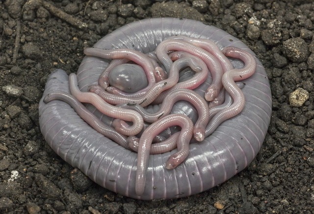
Ringed caecilian feeds milk to her young

Mammals suckle their young. This behaviour distinguishes them from other vertebrates: fish, amphibians, reptiles, and birds. Yet that distinction is not watertight, as a few bird species exist that produce a kind of milk to feed their young: some pigeons and doves, some flamingos, and the emperor penguin. And now, Pedro Mailho-Fontana and colleagues report that females of the ringed caecilian, an amphibian, feed their young with something that resembles mammalian milk. Thanks to this ‘milk’, the youngsters grow rapidly.

It is not surprising that this peculiar trait, which is easily observable, is only now coming to light. The biology of caecilians is poorly known because the animals live underground. Caecilians (Gymnophiona) form a third group within the amphibians, next to frogs & toads and salamanders. They have no legs, have reduced eyes, and are blind; they have two tentacles with which they find their way underground.

The caecilian in question here is the ringed caecilian (Siphonops annulatus). It is widespread in South America. The animal grows to over forty centimetres in length. A female lays eggs, on average ten at a time, in an underground round nest chamber. She is a devoted mother: she lays coiled up with the eggs on her body, and when the young have hatched, she stays with them for another two months; then the young are independent. Until that moment, she will not even leave for a brief time to find food for herself.

A female changes colour when she is attending hatchlings. Normally, ringed caecilians are bluish grey, but a mother turns whitish grey. It was already known that this colour is caused by fat droplets accumulating in her epidermis. Every few days, the young are allowed to scrape off that skin; for this purpose, they have spoon-shaped teeth in the lower jaw. They all do at the same time, ferociously; within ten minutes it is over, and peace returns until the mother’s skin is ready for consumption again. This ‘skin feeding’ occurs in more caecilian species.

But ringed caecilians hatchlings also get other food, Mailho-Fontana discovered: milk. He kept a number of animals in the lab and filmed their behaviour.

Hatchlings often assemble near their mother’s rear end, he observed. Further research revealed that glands in the mother’s oviduct walls produce a white, viscous fluid that emerges through the genital opening, the cloaca, several times a day. The stuff is rich in fats and carbohydrates.

The young imbibe it voraciously. This ‘milk’ is a more important source of nutrition than the mother’s skin, and it is thanks to the milk that the young grow very fast, the researchers think. Their weight doubles within a week after hatching. The mother, eating nothing, loses a third of her weight.

The mother releases milk when hatchlings touch her tail, often producing high-pitched sounds, which probably is begging behaviour. She then raises her body end vertically, and the hatchlings compete for a good place. On average, three of them drink at the same time until they are fully satiated.

Viviparous caecilian species in which the young drink milk in the oviducts before birth were already known. The discovery of oviductal milk in the egg-laying ringed caecilian was unexpected.

Willy van Strien

Photo: Ringed caecilian, female with young. ©Carlos Jared

Sources:
Mailho-Fontana, P.L., M.M. Antoniazzi, G.R. Coelho, D.C. Pimenta, L.P. Fernandes, A. Kupfer, E.D. Brodie Jr. & C. Jared, 2024. Milk provisioning in oviparous caecilian amphibians. Science 383: 1092-1095. Doi: 10.1126/science.adi5379
Jared, C., P.L. Mailho-Fontana, S.G.S. Jared, A. Kupfer, J.H.C. Delabie, M. Wilkinson & M.M. Antoniazzi, 2019. Life history and reproduction of the neotropical caecilian Siphonops annulatus (Amphibia, Gymnophiona, Siphonopidae), with special emphasis on parental care. Acta Zoologica. 100: 292-302. Doi: 10.1111/azo.12254
Wilkinson, M., A. Kupfer, R. Marques-Porto, H. Jeffkins, M.M. Antoniazzi & C. Jared, 2008. One hundred million years of skin feeding? Extended parental care in a Neotropical caecilian (Amphibia: Gymnophiona). Biology Letters 4: 358-361. Doi: 10.1098/rsbl.2008.0217
Repurposed

Tree fern Cyathea rojasiana transforms dead leaves into roots

The tree fern Cyathea rojasiana, which grows in Panama, has a crown of leaves on a trunk. Sometimes a new frond sprouts, sometimes an old leaf dies. But dying is not the end for a leaf, James Dalling and colleagues discovered: the decayed leaf gets a second life. At least: the rachis.

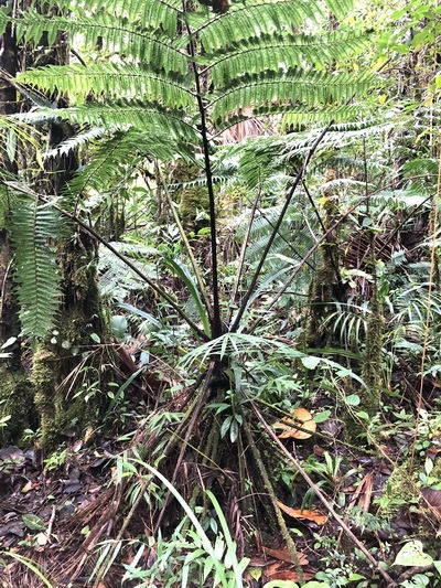
The tree fern grows to a height of two meters, the leaves are more than two meters long. A senescent leaf will bend down, the leaf tip touching the ground. The leaf rots away, but the rachis remains. A tree can have a ‘skirt’ of twenty to thirty of those remains. They look lifeless, but when Dalling tried to remove them, they were found to be firmly stuck in the ground.

Vascular bundles run through the rachis of a green leaf, transporting water and nutrients absorbed by the roots to the leaf tissue. These vascular bundles appear to be intact in the ground-stuck leaf remains of Cyathea rojasiana. They are surrounded by a black layer that apparently protects them from rotting. And, surprisingly, at the end, i.e., in the soil, a bunch of finely branched roots has sprouted from each vascular bundle.

The conclusion is that the former leaf rachises have been transformed into roots, in which the direction of the water flow is reversed: in green leaves, it flowed from stem to leaf tip, now it is from leaf tip to stem.

Cyathea rojasiana grows in wet, extremely nutrient-poor soil. Extra roots are not so much useful to absorb water, the researchers think, but to extract nutrients from a larger area of soil. They showed that the new roots indeed absorb nitrogen and transport it upwards.

For many plant species holds: you can stick a piece of stem or leaf in the ground, and roots will grow. But converting a leaf rachis into a root is something that, as far as we know, only Cyathea rojasiana does.

Willy van Strien

Photo: Tree fern Cyathea rojasiana with decayed leaves, now functioning as roots. ©James Dalling

James Dalling tells about his discovery on YouTube

Source:
Dalling, J.W., E. Garcia, C. Espinosa, C. Pizano. A. Ferrer & J.L. Viana, 2024. Zombie leaves: novel repurposing of senescent fronds in the tree fern Cyathea rojasiana in a tropical montane forest. Ecology e4248, 18 January online. Doi: 10.1002/ecy.4248

Fairy lantern rediscovered

Unexpectedly, the cheating plant Thismia kobensis still exists

The rediscoverd Kobe fariry lantern is a cheater

It was discovered in 1992 and believed to be extinct because the site where it had been found was destroyed in 1999. But now, it is rediscovered elsewhere: the Kobe fairy lantern. Kenji Suetsugu and colleagues describe the beautiful but cheating tiny plant.

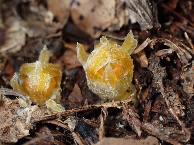
You would hardly recognize them as plants, the small, splendid ‘fairy lanterns’ on the forest floor, often hidden under fallen tree leaves. Fairy lanterns, Thismia species, are indeed remarkable plants. What you see are the flowers, less than a centimeter in size. The plants have no green leaves, only some scales on the very short stem. Most of the plants lives underground.

There are about 90 species, one of which is Thismia kobensis, the Kobe fairy lantern. Small and inconspicuous as it is, it was only discovered in 1992, in an oak forest near the Japanese city of Kobe. The find was small: it consisted of no more than one specimen. The site was destroyed in 1999 when an industrial complex was constructed, and the newly discovered species went extinct. That was what people thought. But fairy tales exist: in 2021 biologists unexpectedly rediscovered the plant on a conifer plantation in the town of Sanda, 30 kilometers from the original site. And this time the find was larger: almost 20 individuals. Now, Kenji Suetsugu and colleagues provide a scientific description of the species.

The loveliness of its flower is deceptive: Thismia kobensis belongs to a group of cheating plants.

Energy requirement

The cheating has to do with the lack of green leaves.

The green leaves of normal plants contain many chloroplasts. In these cellular organelles, photosynthesis takes place: plants extract carbon dioxide from the atmosphere and with the help of sunlight they fix the carbon in carbohydrates such as sugars and starch. From these carbohydrates, they derive energy. Plants without green leaves cannot make carbohydrates, but they do need energy.

Many of these plants solve this problem by extracting sugars with their roots from fungi in the soil. The scientific term for this is mycoheterotrophy.

Fairy lantern is sugar thief

Most mycoheterotrophic plants target fungi that live in a mutualistic relationship with green plants. The fungi get sugars from these plants. In return, the fungi help the green plants to absorb water and nutrients such as nitrogen and phosphorus from the soil. This collaboration, called mycorrhiza, is mutually beneficial and both parties are honest.

However, when mycoheterotrophic plants such as Thismia make contact with mycorrhizal fungi, they don’t cooperate in this way. They do receive water and nutrients, but they do not return sugars. They can’t. Instead, they take up sugars from the fungus in addition to water and nutrients. In other words: they steal. The fungus had received those sugars from green plants, so mycoheterotrophic plants indirectly parasitize on green plants via mycorrhizal fungi.

Difficult alternative

There are about 500 species of mycoheterotrophic plants. They live on nutrient-poor soils in forests, where little sunlight reaches the soil and the ability for photosynthesis, i.e., sugar production, is limited. Sugar theft is the alternative that these plants developed.

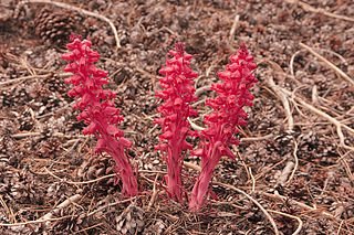
sarcodes sanguinea is a myceheterotrophic plantBut it’s not as easy as it seems. It is difficult for a mycoheterotrophic plant to form a relationship with a mycorrhizal fungus. Where a green plant interacts with many mycorrhizal fungi species simultaneously, a mycoheterotrophic plant can make contact with only one or a few fungal species. That’s probably because most fungi detect the cheaters and hold off on the relationship. Therefore, mycoheterotrophic plants are always rare and never widely distributed.

Mycoheterotrophic species often target a fungus that has many different green partners. With so many suppliers, the sugar supply is always guaranteed.

Dust seeds

The vast majority of land plants live in association with mycorrhizal fungi. The mycoheterotrophic mode of life -which abuses this mutualism – has developed dozens of times. In the case of fairy lanterns, this happened many millions of years ago. That is why they have little resemblance to ordinary plants. Other mycoheterotrophic plants emerged much more recently and have a more normal appearance.

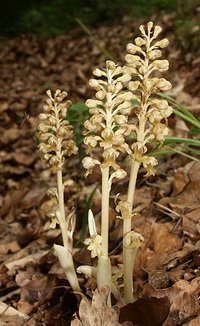
the brid's nest is a mycoheterotrophic orchidSome plants are mycoheterotrophic shortly after germination only; this applies to all orchid species. The seeds are as fine as dust and contain no food. After germination, these plants get their sugars from fungi until they have leaves and can make their own sugars. This could be a first step towards a fully mycoheterotrophic lifestyle. There are also orchid species that stay mycoheterotrophic during their whole life, for example the bird’s nest, Neottia nidus-avis.

Broomrape species (Orobanche) look similar to some mycoheterotrophic plants, but are different: with their roots, they parasitize directly on other plants.

Willy van Strien

Photos:
Large:
Fairy lantern of Kobe, Thismia kobensis ©Kenji Suetsugu
Small:
Snow plant, Sarcodes sanguinea, a mycoheterotrophic plant from North-west America. David῀O (Wikimedia Commons, Creative Commons CC BY 2.0)
Bird’s nest orchid, Neottia nidus-avis. BerndH (Wikimedia Commons, Creative Commons CC BY-SA 3.0)

Sources:
Suetsugu, K., K. Yamana & H. Okada, 2023. Rediscovery of the presumably extinct fairy lantern Thismia kobensis (Thismiaceae) in Hyogo Prefecture, Japan, with discussions on its taxonomy, evolutionary history, and conservation. Phytotaxa 585: 102-112. Doi: 10.11646/phytotaxa.585.2.2
Gomes, S.I.F., M.A. Fortuna, J. Bascompte & V.S.F.T. Merckx, 2022. Mycoheterotrophic plants preferentially target arbuscular mycorrhizal fungi that are highly connected to autotrophic plants. New Phytologist 235: 2034-2045. Doi: 10.1111/nph.18310
Jacquemyn, H. & V.S.F.T. Merckx, 2019. Mycorrhizal symbioses and the evolution of trophic modes in plants. Journal of Ecology 107: 1567-1581. Doi: 10.1111/1365-2745.13165
Gomes, S.I.F., J. Aguirre-Gutiérrez, M.I. Bidartondo & V.S.F.T. Merckx, 2017. Arbuscular mycorrhizal interactions of mycoheterotrophic Thismia are more specialized than in autotrophic plants. New Phytologist 213: 1418-1427. Doi: 10.1111/nph.14249

Incubator

Ants translocate their larvae and pupae to a warm bird’s nest

Myrmica ruginodis translocates brood to a wood warbler's nest

In the nest of a wood warbler not only young wood warblers may grow up, but also ants, as Marta Maziarz and colleagues discovered. Ant larvae and pupae probably survive, grow and develop better in the bird’s nest than in their own nest.

Usually, nests of European forest ants, especially Myrmica ruginodis and Myrmica rubra (the European fire ant or common red ant), are so cold in spring that the larvae and pupae do not grow well. Their development only starts at 16°C, but ant nests rarely get that warm before summer. They are located on the forest floor, between fallen leaves of deciduous trees. The ants cannot produce heat, so without sunlight the nests have the same temperature as the environment. A temperature of 20 to 25°C is optimal for raising brood; the nests never reach that temperature in spring.

But warm places are available nearby, Marta Maziarz and colleagues show. On the forest floor, the wood warbler, a songbird that breeds in European forests, makes a domed nest of grasses, leaves and moss. The bodies of bird parents and, later on, their fully feathered young keep the nest warm.

Translocation

When the bird parents incubate the eggs, in the second half of May, the nest temperature often reaches 16°C or more; especially on cold days, the difference with the ambient temperature is large. Once the young birds are fully feathered, in the first week of June, the nest temperature even rises to 20°C and higher.

This is a nice temperature for the ants, which indeed visit the warm wood warbler’s nest. In cold weather in May and in the first week of June, they move larvae and pupae from their own nest to a bird’s nest and put them in the sidewalls. Translocation is a lot of work, but apparently, it is worth the effort.

After fledging, the vacant nest cools down again. But the ants don’t remove their brood immediately; they delay relocation for up to two weeks. There is no hurry, because the bird’s nest is no longer warm, but it is not colder than the ants’ nest either.

In the primeval forest of Białowieża in Poland, where Maziarz conducted the research, the ants use 10 to 30 percent of wood warblers’ nests as incubators. The birds do not suffer from the inhabitants, but they do not benefit from them either. Therefore, they do not specifically seek the proximity of ant colonies when starting nest building. It is the ants that start the relationship and benefit from it.

Willy van Strien

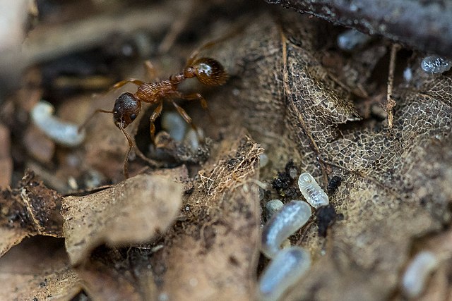
Photo: Myrmica ruginodis with brood. Jan Anskeit (Wikimedia Commons, Creative Commons CC BY 4.0)

Sources:
Maziarz, M., R.K. Broughton, L.P. Casacci, G. Hebda, I. Maak, G. Trigos‑Peral & M. Witek, 2021. Interspecific attraction between ground‑nesting songbirds and ants: the role of nest‑site selection. Frontiers in Zoology 18: 43. Doi: 10.1186/s12983-021-00429-6
Maziarz, M., R.K. Broughton, L.P. Casacci, A. Dubiec, I. Maák & M. Witek, 2020. Thermal ecosystem engineering by songbirds promotes a symbiotic relationship with ants. Scientific Reports 10: 20330. Doi: 10.1038/s41598-020-77360-z

New body

Loose head regenerates a complete Elysia sea slug

Elysia sea slug can grow new body from head

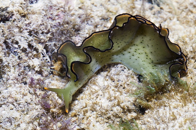
Sea slugs Elysia marginata en Elysia atroviridis can decapitate themselves and regrow a new body from the loose head, Sayaka Mitoh en Yoichi Yusa show. A bizarre phenomenon. Why do they do it, and how do they survive?

Sayaka Mitoh and Yoichi Yusa must have been dumbfounded when seeing sea slugs that they kept in their lab, species Elysia marginata, sever their heads from their bodies. The loose heads moved around, as they report. After a day, the wounds were closed. In some cases, especially in young sea slugs, things got even crazier: the head began to feed; after a week, a new body started to grow and in three weeks it was complete.

The loose bodies also moved for a while, sometimes even months, but eventually they decomposed. No new head appeared on any loose body.

Parasite

There are more animals that can regrow a missing body part, such as a lizard that shed its tail or a fiddle crab that lost a claw. But this – regenerating almost an entire body – is very extreme. These sea slugs even have a groove behind the head as a predetermined breakage plane for self-decapitation. Why do they do it?

In any case, it is not to escape from a predator, like a lizard sheds its tail when a predator grasps it. The sea slugs take hours to separate body from head; that is not effective to avoid predation. And when the researchers simulated an attack by teasing them, nothing happened. The animals have a different defence mechanism against predators: they are poisonous.

The reason for self-decapitation became clear by observations on wild-caught specimens of a related species, Elysia atroviridis. Once in the lab, some of them shed the whole body, and these specimens turned out to contain a parasite, a copepod of the genus Arthurius. It is a large parasite that occupies almost the entire body of its host. In fact, a parasitized sea slug has already lost its body. If it sheds it, it will get rid of the parasite while losing almost nothing more.

Chloroplasts

But how does it survive without organs such as heart and kidneys? This has to do with a special property of sacoglossan sea slugs, to which Elysia belongs, the researchers suppose. They extract chloroplasts from algal food and incorporate them in special cells that line their highly branched digestive gland. The head also contains chloroplasts. Thanks to the chloroplasts, which they need to survive, these sea slugs can endure a period without food, it was known.

It is a mystery how exactly they utilise the chloroplasts. The chloroplasts continue to do what they do in plants: they convert carbon dioxide into carbohydrates with the help of sunlight, a process called photosynthesis. Whether the sea slugs can survive on sunlight as a result, just like plants, is a matter of debate.

Regardless, it may well be thanks to the chloroplasts that a loose head of Elysia marginata and Elysia atroviridis survives.

No eternal life

Parasitized Elysia sea slugs shed their worthless bodies. But they only manage to grow a new one from the head when they are young. The loose head of an older specimen does not feed and does not grow, but will die within ten days. Shedding and regrowing a body is not a recipe for eternal life.

Willy van Strien

Photo: Elysia marginata. Budak (via Flickr, CC BY-NC-ND 2.0)

The research explained on YouTube

Sources:
Mitoh, S. & Y. Yusa, 2021. Extreme autotomy and whole-body regeneration in photosynthetic sea slugs. Current Biology 31: R233-R234. Doi: 10.1016/j.cub.2021.01.014
Wägele, H., 2015. Photosynthesis and the role of plastids (kleptoplastids) in Sacoglossa (Heterobranchia, Gastropoda): a short review. Aquatic Science & Management 3: 1-7. Doi: 10.35800/jasm.3.1.2015.12431

Collateral benefit

Bird disperses eggs of stick insects it swallowed

brown-eared bulbul disperses eggs of stick insects

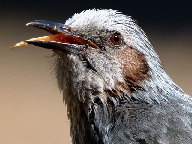
Some stick insects are even more like plants than you might think at first glance. Just like plant seeds, the eggs can be dispersed by a bird, Kenji Suetsugu and colleagues show.

Stick insects are perfectly camouflaged: they do not stand out among the plants. Yet insect-eating birds are able to find them and will eat them. And that is the end of the story for such tiny animal.

Well, it may not be, Kenji Suetsugu and colleagues report. If an unfortunate female stick insect is carrying mature eggs, a few of these appear undamaged in the bird’s droppings, and some may even hatch.

Younsters

The researchers, working in Japan, point out that the eggs of stick insects resemble plant seeds: they have the same size and colour and feel the same thanks to a hard shell. Hence their suggestion that the eggs might survive passage through a bird’s digestive tract like plant seeds do. Many plant species produce fruits that are eaten by birds or other animals; the seeds remain intact, are excreted and germinate. Is something similar possible for the eggs of stick insects?

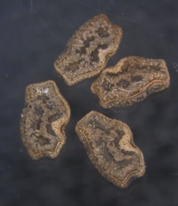
eggs of stick insect after passage through a bird's digestive tractTo find out, they mixed mature eggs of three stick insect species with an artificial diet and fed this to a brown-ear bulbul, one of the main predators of the insects. Afterwards, they examined the bird’s droppings under a stereomicroscope and discovered a small number of intact eggs, and from some of these eggs a young stick insect hatched later on.

Such scenario is also possible when a bird swallowed a gravid female, the authors think. The youngsters that hatch after passage through a bird’s guts would have to find an appropriate food plant to live on, but that is always the case. Normally, a female just drops her eggs to the ground and does not provide any care.

Parthenogenetic

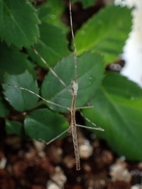
young stick insect, hatched from egg that passed through a bird's gutsSo, sick insects not only look like plants, but they also exhibit a surprising plant-like trait: dispersal of offspring by birds, which is unique in insects.

Dispersal by an avian predator is only possible for species that reproduce parthenogenetically, for in that case females carry eggs that can develop without fertilization. A number of stick insect species exhibit parthenogenesis, including the species that were studied here.

Flyways

Dispersal of insect eggs via a bird’s digestive tract is not entirely comparable to dispersal of plant seeds. Plants produce fruits that have to be eaten to disperse their seeds. In contrast, a female stick insect has no intention to be captured by a bird to have her eggs transported – by being camouflaged, she tries to prevent just that. But if she is unlucky enough to become a bird’s meal, it is a collateral benefit if some eggs survive and young hatch, if only a few.

The hard eggs probably have not evolved to facilitate avian dispersal, the authors suggest, but to decrease the risk of attack by parasitoid wasps, which lay their eggs in other insects’ eggs.

Stick insects are immobile. Thanks to the birds they may reach new places to live. An interesting question is whether distribution patterns in the insects, to be unravelled by DNA research, overlap with birds’ flyways; that would strengthen the idea that the eggs are sometimes dispersed like plant seeds.

Willy van Strien

Photos
Large: Brown-eared bulbul (tongue visible). Alpsdake (Wikimedia Commons, Creative Commons BY-SA 4.0)
Small: stick insect (Ramulus irregulariterdentatus) eggs that passed through a bird’s digestive tract and a young stick insect that hatched from such egg. ©Kenji Suetsugu

Source:
Suetsugu, K., S. Funaki, A. Takahashi, K. Ito & T. Yokoyama, 2018. Potential role of bird predation in the dispersal of otherwise flightless stick insects. Ecology, online May 29. Doi: 10.002/ecy.2230

Nutritious two-component glue

Queen larva is firmly attached to her ceiling

Royal jelly, fed to a queen larva, holds her in place

A bee larva that is to become a queen receives large quantities of royal jelly. And that is not only because the stuff is nutritious, as Anja Buttstedt and colleagues show.

A female honeybee larva can become a worker or a queen, her fate depending on the food she receives. During the first days, all larvae are treated to the so-called royal jelly, a nutritious mixture that the nurse bees produce in their head glands; it is rich in proteins, sugars and fats. After the third day, larvae that will grow up to be worker bees are raised on a different diet. When they pupate, nurse bees close their cells with a layer of wax. But a larva that is destined to become a queen is fed on royal jelly exclusively; the nurse bees bring it to her in generous quantities. Thanks to that nutritious diet, she grows bigger than worker bees.

Queen cup

The royal jelly has still another function, Anja Buttstedt and colleagues discovered: it holds the queen larva in place.

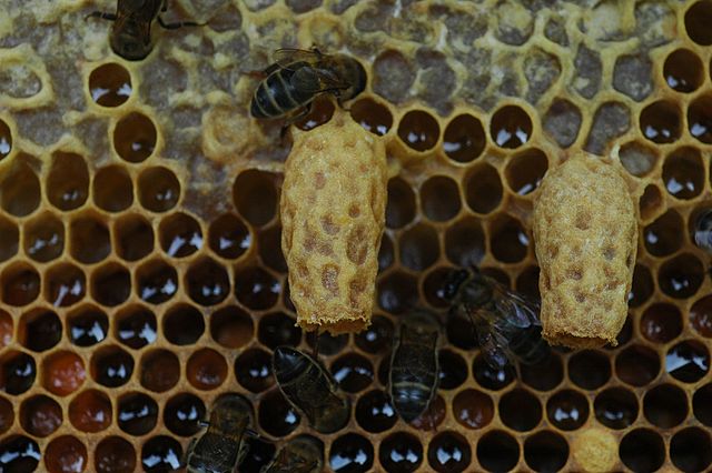
And that is badly needed. The cells in the comb, in which worker larvae grow up, are too small for a developing queen larva. For her, the bee workers will build a special cell, a so-called queen cell or queen cup. It is not only wider, but also differently oriented: vertically, opening downwards. Therefore, her royal highness could easily fall out of her cell.

Buttstedt shows why that does not happen: the royal jelly, which the workers deposit on the ceiling, is so sticky that it keeps the larva hanging from the ceiling until it pupates and the cell is sealed with wax. The stickiness arises because two proteins, royalactin (the main protein in royal jelly) and apisimin, form long fibrous structures that make the jelly viscous.

Fiber network

The workers produce and store the proteins in their hypopharyngeal glands. The gland mixture is liquid, enabling the bees to excrete it. But when they deposit it in a brood cell, they combine it with fatty acids which they produced in the mandibular glands, and in those acidic conditions, the proteins royalactin and apisimin form a fiber network.

So, royal jelly is a two-component adhesive, as the authors conclude, serving as excellent food as well. It is just what a queen larva needs to grow up safely.

Willy van Strien

Photo: Honeybee, comb and two queen cells. Piscisgate (Wikimedia Commons, Creative Commons CC BY-SA 4.0)

Source:
Buttstedt, A., C.I. Muresxan,H. Lilie, G. Hause, C.H. Ihling, S-H. Schulze, M. Pietzsch & R.F.A. Moritz, 2018. How honeybees defy gravity with royal jelly to raise queens. Current Biology, online March 15. Doi: 10.1016/j.cub.2018.02.022