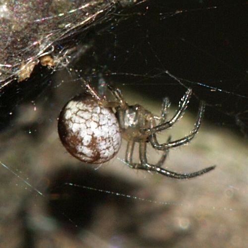
From so simple a beginning

Evolution and Biodiversity

Page 3 of 19

Wet plumage

Namaqua sandgrouse father carries water for the chicks

Male Namaqua sandgrouse fetches water for his chicks with specially adapted belly feathers

As long as the chicks are unable to fly, a Namaqua sandgrouse father will fetch water for them. Jochen Mueller and Lorna Gibson describe the specially adapted belly feathers that enable this.

As their name suggests, sandgrouse species live in dry, almost barren places. The Namaqua sandgrouse (Pterocles namaqua), for example, lives in deserts in Southwest Africa, such as the Kalahari and the Namibian desert. The birds breed up to no less than 30 kilometers from the nearest body of water. Because they mainly eat dry seeds, they have to drink. Adult birds therefore fly to waterholes in the morning and evening. This is how they survive in their arid habitat.

But their chicks can’t go with them to the waterholes for the first month. They are immediately independent after hatching; they walk and forage for food on their own. But they can’t fly yet. It was already known that sandgrouse fathers transport a supply of water for the young in specially adapted belly feathers, which trap and hold water. Now, using various microscopic techniques, Jochen Mueller and Lorna Gibson describe the structure of those feathers in detail, both in wet and dry state.

Fringe

To stock up a supply of water, a Namaqua sandgrouse male steps into the water until it reaches his belly. He fluffs up his belly feathers and rocks his body, soaking the feathers. Then, he presses his belly feathers against his body and leaves. He can store an estimated 25 milliliters of water, 15 percent of his body weight. He flies back at high speed, a trip that can take half an hour. During the flight through dry desert air, some water evaporates, but a lot is still left when he arrives at his nest.

The chicks run up to him and strip the wet feathers with their beaks.

That the belly feathers have a special structure, can already be seen with the naked eye. The feathers have a broad hairy fringe along the side, except at the top. But only under the microscope does the special structure reveal itself completely.

Coiled barbules

A normal bird’s contour feather consists of a shaft on which barbs are implanted, from which barbules branch. These barbules interlock with hooklets and grooves, giving the feather a closed plane. Thanks to the hooklets and grooves, a crumpled feather can be rubbed back into shape.

Under the microscope, the barbs and barbules of the belly feathers of Namaqua sandgrouse males appear to have a different structure. The hairy fringe along the feather is formed by the outer part of the barbs being thin and flexible, and the barbules implanted on the outer part being thin and flexible too.

The inner part of the barbs, where they are attached to the shaft up to just over mid-length, is thicker and stiff. The barbules on this part branch at the upper side, make one helical curl downward and straighten out, running parallel to the barb. The coils of successive barbules intertwine and keep the feather surface closed.

That is how a belly feather looks when it is dry.

Storing water

If such feather gets wet, the picture changes. The barbules on the inner part of the barb uncurl and bend downwards perpendicularly to the feather plane, forming a dense forest of fibers. Due to the so-called capillary force, water is sucked up and held between them.

The fringe of the feather (i.e., the outer parts of the barbs and the barbules that branch from those parts) bends down and inward to the feather shaft, creating a layer to hold the water.

The Namaqua sandgrouse is one of 14 species of sandgrouse (Pterocles), all of which live on arid terrain. In all these species, the males can carry water in their belly feathers, thanks to that unique adaptation of the feather structure.

Willy van Strien

Photo: Pterocles namaqua, male. Bernard DUPONT (Wikimedia Commons, Creative Commons CC BY-SA 2.0)

On YouTube: Namaqua sandgrouse male fetches water for chicks

Sources:
Mueller, J. & L.J. Gibson, 2023. Structure and mechanics of water-holding feathers of Namaqua sandgrouse (Pterocles namaqua). Journal of the Royal Society Interface 20: 20220878. Doi: 10.1098/rsif.2022.0878
Cade, T.J. & G.L. Maclean, 1967. Transport of water by adult sandgrouse to their young. The Condor 69: 323-343. Doi: 10.2307/1366197

Reinforced carton

Crematogaster clariventris grows a fungus that strengthens its nest wall

Crematogaster clariventris grows a fungus that reinforces its nest

Workers of the ant Crematogaster clariventris collect pieces of fresh leaves to grow a fungus, Alain Dejean and colleagues observed. The threads of the fungus reinforce the carton nest of the ants.

Fungus threads as a component of building or insulation material: you hear more and more about it. It is considered to be innovative, but……. ants were ahead of us. Some species strengthen the walls of their nests with fungal hyphae (threads). The African ant Crematogaster clariventris even collects fresh pieces of leaves to feed them to a fungus that forms strong hyphae, Alain Dejean and colleagues discovered.

The ant lives in large colonies, high in trees. On main branches, workers build nests of hard carton, which they make by chewing fibrous plant material, such as hairs (trichomes) or pieces of wood. They add a fungus, with the result that a network of branched fungal hyphae is embedded in the carton walls; the hyphae consist of tubular cells with a sturdy cell wall. The nest wall is a natural composite material.

Fresh leaf

Dejean, who works in Cameroon, noticed that workers of Crematogaster clariventris bring freshly cut pieces of young and nutritious plant leaves whenever a new nest is constructed or a damaged part of a nest is repaired. Other workers add chewed pulp, and the whole hardens into fungus-reinforced carton in a few days. From these observations, the researchers deduce that the ants bring the fresh leaf material as food for the fungus that forms reinforcing hyphae, so that it will grow well in the new nest wall.

After the fungus died, the sturdy hyphae in the nest wall remain intact.

Crematogaster clariventris is not the only ant species to cut off pieces of leaves to grow a fungus. In Central and South America, ant species occur that cut pieces of fresh leaves and carry it to fungus gardens in their underground nests, the leafcutter ants. They grow fungus for food. So, ants also preceded us in agriculture.

Willy van Strien

Photo: Crematogaster clariventris ©Piotr Naskrecki

Source:
Dejean, A., P. Naskrecki, C. Faucher, F. Azémar, M. Tindo, S. Manzi & H. Gryta, 2023. An Old World leaf-cutting, fungus-growing ant: A case of convergent evolution Ecology & Evolution 13: e9904. Doi: 10.1002/ece3.9904

Super white

Woodcock feathers have the whitest white of all birds

Tail feathers of woodcock are brilliant white at the underside

The whitest feathers that exist can be found in the woodcock, which otherwise has an inconspicuous appearance. Jamie Dunning and colleagues investigated how the surprisingly white hue emerges.

An Eurasian woodcock (Scolopax rusticola) is so well camouflaged that it hardly stands out against the forest floor on which it lives. But the tips of its tail feathers are brilliant white on the underside and therefore very visible, even in dim light. No plumage exist with patches that are whiter than those feather tips. Jamie Dunning and colleagues show how that super white hue is brought about by the structure of the tail feathers.

Woodcocks rest during the day, and then it is important not to stand out. Hence their mottled brown plumage. At dawn or dusk, they are active. To show themselves to each other, they raise their short tails or make a courtship flight. Then, the bright white tips on the underside of the tail feathers stand out clearly.

Nanostructure

Those white tail tips are conspicuous at dim light because they reflect much of the scarce light that falls on them. This is possible because of a special structure. A bird’s feather consists of a shaft on which barbs are implanted. The barbs of the super-white feather tips of Eurasian woodcocks are flattened and thickened, and, like the slats of Venetian blinds, they are slanted and overlap. As a result, a maximal amount of light is reflected.

But before the light rays bounce back, they are scattered beneath the surface of the barbs. The barbs have a disordered internal structure of nanofibers and scattered air pockets, which causes incident light rays to change direction frequently and chaotically. This strong so-called diffuse reflection results in a bright white appearance, just as happens in snow.

The barbs are held together by the many Velcro-like barbules that branch from them. These are brownish, but because they are on the upper side of the tail feathers, they do not affect the whiteness of the underside.

The Eurasian woodcock lives in Europe and Asia. There are seven other woodcock species worldwide, all with super white tops at the underside of the tail feathers. Other birds don’t possess such white feather patches, not even species that are closely related to woodcocks, such as common snipe (Gallinago gallinago).

Willy van Strien

Photo: American woodcock, Scolopax minor, with raised tail. Matt Schenck (Wikimedia Commons, Creative Commons CC BY 4.0)

See also: super black feathers also exist

Source:
Dunning, J., A. Patil, L. D’Alba, A.L. Bond, G. Debruyn, A. Dhinojwala, M. Shawkey & L. Jenni, 2023. How woodcocks produce the most brilliant white plumage patches among the birds. Interface 20: 20220920. Doi: 10.1098/rsif.2022.0920

Fairy lantern rediscovered

Unexpectedly, the cheating plant Thismia kobensis still exists

The rediscoverd Kobe fariry lantern is a cheater

It was discovered in 1992 and believed to be extinct because the site where it had been found was destroyed in 1999. But now, it is rediscovered elsewhere: the Kobe fairy lantern. Kenji Suetsugu and colleagues describe the beautiful but cheating tiny plant.

You would hardly recognize them as plants, the small, splendid ‘fairy lanterns’ on the forest floor, often hidden under fallen tree leaves. Fairy lanterns, Thismia species, are indeed remarkable plants. What you see are the flowers, less than a centimeter in size. The plants have no green leaves, only some scales on the very short stem. Most of the plants lives underground.

There are about 90 species, one of which is Thismia kobensis, the Kobe fairy lantern. Small and inconspicuous as it is, it was only discovered in 1992, in an oak forest near the Japanese city of Kobe. The find was small: it consisted of no more than one specimen. The site was destroyed in 1999 when an industrial complex was constructed, and the newly discovered species went extinct. That was what people thought. But fairy tales exist: in 2021 biologists unexpectedly rediscovered the plant on a conifer plantation in the town of Sanda, 30 kilometers from the original site. And this time the find was larger: almost 20 individuals. Now, Kenji Suetsugu and colleagues provide a scientific description of the species.

The loveliness of its flower is deceptive: Thismia kobensis belongs to a group of cheating plants.

Energy requirement

The cheating has to do with the lack of green leaves.

The green leaves of normal plants contain many chloroplasts. In these cellular organelles, photosynthesis takes place: plants extract carbon dioxide from the atmosphere and with the help of sunlight they fix the carbon in carbohydrates such as sugars and starch. From these carbohydrates, they derive energy. Plants without green leaves cannot make carbohydrates, but they do need energy.

Many of these plants solve this problem by extracting sugars with their roots from fungi in the soil. The scientific term for this is mycoheterotrophy.

Fairy lantern is sugar thief

Most mycoheterotrophic plants target fungi that live in a mutualistic relationship with green plants. The fungi get sugars from these plants. In return, the fungi help the green plants to absorb water and nutrients such as nitrogen and phosphorus from the soil. This collaboration, called mycorrhiza, is mutually beneficial and both parties are honest.

However, when mycoheterotrophic plants such as Thismia make contact with mycorrhizal fungi, they don’t cooperate in this way. They do receive water and nutrients, but they do not return sugars. They can’t. Instead, they take up sugars from the fungus in addition to water and nutrients. In other words: they steal. The fungus had received those sugars from green plants, so mycoheterotrophic plants indirectly parasitize on green plants via mycorrhizal fungi.

Difficult alternative

There are about 500 species of mycoheterotrophic plants. They live on nutrient-poor soils in forests, where little sunlight reaches the soil and the ability for photosynthesis, i.e., sugar production, is limited. Sugar theft is the alternative that these plants developed.

sarcodes sanguinea is a myceheterotrophic plantBut it’s not as easy as it seems. It is difficult for a mycoheterotrophic plant to form a relationship with a mycorrhizal fungus. Where a green plant interacts with many mycorrhizal fungi species simultaneously, a mycoheterotrophic plant can make contact with only one or a few fungal species. That’s probably because most fungi detect the cheaters and hold off on the relationship. Therefore, mycoheterotrophic plants are always rare and never widely distributed.

Mycoheterotrophic species often target a fungus that has many different green partners. With so many suppliers, the sugar supply is always guaranteed.

Dust seeds

The vast majority of land plants live in association with mycorrhizal fungi. The mycoheterotrophic mode of life -which abuses this mutualism – has developed dozens of times. In the case of fairy lanterns, this happened many millions of years ago. That is why they have little resemblance to ordinary plants. Other mycoheterotrophic plants emerged much more recently and have a more normal appearance.

the brid's nest is a mycoheterotrophic orchidSome plants are mycoheterotrophic shortly after germination only; this applies to all orchid species. The seeds are as fine as dust and contain no food. After germination, these plants get their sugars from fungi until they have leaves and can make their own sugars. This could be a first step towards a fully mycoheterotrophic lifestyle. There are also orchid species that stay mycoheterotrophic during their whole life, for example the bird’s nest, Neottia nidus-avis.

Broomrape species (Orobanche) look similar to some mycoheterotrophic plants, but are different: with their roots, they parasitize directly on other plants.

Willy van Strien

Photos:
Large:
Fairy lantern of Kobe, Thismia kobensis ©Kenji Suetsugu
Small:
Snow plant, Sarcodes sanguinea, a mycoheterotrophic plant from North-west America. David῀O (Wikimedia Commons, Creative Commons CC BY 2.0)
Bird’s nest orchid, Neottia nidus-avis. BerndH (Wikimedia Commons, Creative Commons CC BY-SA 3.0)

Sources:
Suetsugu, K., K. Yamana & H. Okada, 2023. Rediscovery of the presumably extinct fairy lantern Thismia kobensis (Thismiaceae) in Hyogo Prefecture, Japan, with discussions on its taxonomy, evolutionary history, and conservation. Phytotaxa 585: 102-112. Doi: 10.11646/phytotaxa.585.2.2
Gomes, S.I.F., M.A. Fortuna, J. Bascompte & V.S.F.T. Merckx, 2022. Mycoheterotrophic plants preferentially target arbuscular mycorrhizal fungi that are highly connected to autotrophic plants. New Phytologist 235: 2034-2045. Doi: 10.1111/nph.18310
Jacquemyn, H. & V.S.F.T. Merckx, 2019. Mycorrhizal symbioses and the evolution of trophic modes in plants. Journal of Ecology 107: 1567-1581. Doi: 10.1111/1365-2745.13165
Gomes, S.I.F., J. Aguirre-Gutiérrez, M.I. Bidartondo & V.S.F.T. Merckx, 2017. Arbuscular mycorrhizal interactions of mycoheterotrophic Thismia are more specialized than in autotrophic plants. New Phytologist 213: 1418-1427. Doi: 10.1111/nph.14249

Cleaning ants are successful

Metarhizium fungus makes fewer victims

Argentine ant removes sporen of Metarhizium fungus

Ants defend themselves against disease-causing Metarhizium fungus by grooming off fungal spores from each other. Prolonged exposure to that cleaning behaviour makes the fungus less deadly, Miriam Stock and colleagues show.

Metarhizium fungus can quickly spread throughout an ant nest because the ants easily infect each other with fungal spores. But the animals take action to inhibit the pathogen. That does not leave the fungus unaffected, Miriam Stock and colleagues show with experiments.

To counteract the fungus, ants can disinfect nest and brood (eggs, larvae and pupae) with a mixture of formic acid, which they produce in a poison gland, and tree resin. In addition, a sick ant stays away from the brood and spends more and more time outside the nest so as not to endanger its nest-mates. And the animals keep each other clean. If spores of the fungus land on an ant, her nest-mates either groom off the spores, risking infection themselves, or spray them with formic acid.

New spores

These caring nest-mates should act quickly. The spores attach on the affected ant and germinate, after which nothing can be done anymore. The fungus penetrates the body to develop, eventually killing the ant. Then the fungus appears on the cadaver forming spores that make new victims in the next infection cycle.

Conducting experiments with the Argentine ant, Linepithema humile, Stock shows that timely care does indeed help; the presence of other ants reduces the chance that an ant dies after contact with fungal spores.

But, as it turns out, cleaning also causes changes in the fungus.

Metarhizium-fungus adapts

The trials consisted of series in which the Metarhizium fungus passed repeatedly via spores from a dead ant to a new victim. In half of these series, the infected ant was held isolated, in the other half she was accompanied by two nest-mates that could remove the fungal spores. Conducting a final test after ten infection cycles, the researchers allowed the fungus to infect either an isolated ant or an ant with company.

In the final test, fungal lines that had grown on isolated ants caused a lot of mortality among newly infected ants when they did not receive care from others. But fungal lines that had infected ants that were in company of other ants – that could groom them -, had changed. They formed twice as many spores, but nevertheless made fewer victims among ants they came into contact with, even if there were no nest-mates around to help. These fungal lines had become less deadly.

Essential component

And there was something else: the spores of those ‘social fungal lines’ were less well detected and removed by the ants. The researchers discovered that these spores produced less ergosterol; this is a compound that occurs in all fungi and that, apparently, arouses the ants. So, the ‘social fungus lines’ evade defence by the ants.

But this comes at a cost. Ergosterol is an essential component of the spore membrane. The fact that the ‘social lines’ have lower levels of this important component probably explains why they are less deadly.

So, grooming each other to remove Metarhizium fungus spores as ants do is useful in two ways. It works immediately if ants quickly remove spores from a nest-mate, saving her from death. And in the longer term, it makes the fungus less dangerous.

Willy van Strien

Photo: Argentine ants exchanging food. Davefoc (Wikimedia Commons, Creative Commons CC BY-SA 4.0)

See also: ants disinfect their nest with a mixture of resin and formic acid

Sources:
Stock, M., B. Milutinović, M. Hoenigsberger, A.V. Grasse, F. Wiesenhofer, N. Kampleitner, M. Narasimhan, T. Schmitt & S. Cremer, 2023. Pathogen evasion of social immunity. Nature Ecology & Evolution, online February 2. Doi: 10.1038/s41559-023-01981-6
Brütsch, T., G. Jaffuel, A. Vallat, T.C.J. Turlings & M. Chapuisat, 2017. Wood ants produce a potent antimicrobial agent by applying formic acid on tree-collected resin. Ecology and Evolution 7: 2249-2254. Doi: 10.1002/ece3.2834
Bos, N., T. Lefèvre, A.B. Jensen & P. D’Ettore, 2012. Sick ants become unsociable. Journal of Evolutionary Biology 25: 342-351. Doi: 10.1111/j.1420-9101.2011.02425.x
Chapuisat, M., A. Oppliger, P. Magliano & P. Christe, 2007. Wood ants use resin to protect themselves against pathogens. Proceedings of the Royal Society B 274: 2013-2017. Doi: 10.1098/rspb.2007.0531

Red blood cells hided

Glass frog is more translucent when sleeping

Fleischmann's glass frog is extra translucent when sleeping.

A sleeping Fleischmann’s glass frog can hardly be seen. Red blood cells, which would make the animal visible, are stored away temporarily, Carlos Taboada and colleagues write.

Fleischmann’s glass frog has transparent muscles and a transparent ventral skin that transmit light, rendering heart and intestines visible from below. The skin of its back contains a little green pigment. With these qualities, the animal is translucent: a form of camouflage. But red blood cells – which do not transmit the light, but reflect red light and absorb other colours – can spoil the effect.

Carlos Taboada and colleagues show that the frog has a way to solve this problem: when it sleeps, it removes almost all red blood cells from the bloodstream.

Sleep during daytime

Glass frogs belong to the few translucent land animals that exist. Fleischmann’s glass frog, Hyalinobatrachium fleischmanni, is one of them. The animal, which grows up to three centimetres in length, is found in rainforests in Central and South America. Adult frogs live on land. They are active at night and sleep during daytime, hanging upside down under a leaf. The less they stand out against the leaf when sleeping, the harder it is for predators, mainly birds, to spot them.

It is helpfull that the glass frog is translucet. And by removing almost all red blood cells, about 90 percent, from circulation, a sleeping glass makes itself extra translucent. It hides the red blood cells in the liver, which expands considerably as a result. So, the glass frog is more difficult to detect while resting, when it cannot be alert. As soon as the animal resumes activity, the blood cells go back into the bloodstream and translucency diminishes.

Oxygen

Red blood cells are red because they contain the pigment haemoglobin, a protein that binds oxygen; red blood cells carry oxygen to all other cells. During sleep, therefore, the cells receive no oxygen. Apparently, they are able to coop with that.

Willy van Strien

Photo: Fleischmann’s glass frog. Esteban Alzate (Wikimedia Commons, Creative Commons CC BY-SA 2.5)

Sources:
Taboada, C., J. Delia, M. Chen, C. Ma, X. Peng, X. Zhu, L. Jiang, T. Vu, Q. Zhou, J. Yao, L. O’Connell & S. Johnsen, 2022. Glassfrogs conceal blood in their liver to maintain transparency. Science 378: 1315-1320. Doi: 10.1126/science.abl662
Cruz, N.M. & R.M. White, 2022.  Lessons on transparency from the glassfrog. Transparency in glassfrogs has potential implications for human blood clotting. Science 378: 1272-1273. Doi: 10.1126/science.adf75
Barnett, J.B., C. Michalis, H.M. Anderson, B.L. McEwen, J. Yeager, J.N. Pruitt, N.E. Scott-Samuel & I.C. Cuthill, 2020. Imperfect transparency and camouflage in glass frogs. PNAS 117: 12885-12890. Doi: 10.1073/pnas.1919417117

Gaping display

Sarcastic fringehead impresses with giant upper jaw

sarcastic fringehead can open its mouth extraordinary wide

Males of the blenny Neoclinus blanchardi, the sarcastic fringehead, can open their mouths extraordinary wide. They perform their gaping display for nothing but impressing each other, Watcharapong Hongjamrassilp and colleagues show.

It is an amazing scene when Neoclinus blanchardi fully opens its mouth. A huge membrane becomes visible, consisting of palate and cheeks. It is vividly coloured and has a yellow margin.

In English, the fish is called sarcastic fringehead; it lives along the coast of California. It was already known that males, that have a larger mouth than females, impress each other with it. Now, Watcharapong Hongjamrassilp and colleagues show that they perform their elaborate display for that purpose exclusively.

Wrestling

The sarcastic fringehead can open a large mouth thanks to an upper jaw that is enlarged compared to related fish species and that grows longer than the rest of the body. Its posterior part is unossified and flexible and extends beyond the head. The jaw is connected to the skull in such a way that it can be rotated laterally.

As said, males signal by gaping to scare off each other. A successful male manages to occupy an empty snail shell or a rock crevice in which he hides with only his head protruding. He tries to attract a female. When she likes him, she will lay eggs in his shelter. He fertilizes the eggs and takes care of them until the young hatch. When another male invades his territory, he approaches him, performing the gape display.

The intruder either retreats immediately, or he persists and returns the display. Then the males clash and push each other with the open mouths pressed together. The bigger a male is, the wider his gape. The smallest usually loses, sometimes after the victor had bitten him.

Apparently, suitable shelters are so scarce that the fish has developed a special weapon to defend its place.

Missed opportunity?

But the sarcastic fringehead’s exaggerated gape would also be impressive enough to scare away predators, Hongjamrassilp thought. Or enticing enough to seduce females. He scuba-dove into the water and conducted experiments in the laboratory to see whether this happens.

It did not. If a predator looms, the sarcastic fringehead chases it away by burst swimming. And when a female shows up, he rapidly shakes his head side to side to arouse her interest. He keeps his amazing mouth closed in both cases.

A missed opportunity, you might say.

Willy van Strien

Photo: Neoclinus blanchardi in its shelter. Magnus Kjaergaard (Wikimedia Commons, Creative Commons CC BY-SA 2.5)

This video shows the gaping display

Sources:
Hongjamrassilp, W., Z. Skelton & P.A. Hastings, 2022. Function of an extraordinary display in Sarcastic Fringeheads (Neoclinus blanchardi) with comments on its evolution. Ecology, online Octobre 6: e3878. Doi: 10.1002/ecy.3878
Hongjamrassilp, W., A.P. Summers & P.A. Hastings, 2018. Heterochrony in fringeheads (Neoclinus) and amplification of an extraordinary aggressive display in the Sarcastic Fringehead (Teleostei: Blenniiformes). Journal of Morphology 279: 626-635. Doi:10.1002/jmor.20798

Cuckoo duck seeks defence

Foster family protects duck eggs against birds of prey

Cuckoo duck dumps its eggs in nest of aggressive host

Young cuckoo ducks do not need any care: they are independent upon hatching. Then why does the duck burden other birds with its eggs, Bruce Lyon and colleagues wondered.

In South America a duck species occurs that, like a cuckoo, lays its eggs in nests of other bird species. The hosts then unintentionally take care of them. This is the black-headed duck, Heteronetta atricapilla, with the appropriate nickname cuckoo duck; it is a so-called brood parasite.

Bruce Lyon and colleagues wondered why the cuckoo duck dumps its eggs in other birds’ nests. They don’t require much care, apart from brooding. After hatching, the young are immediately independent. That is a big difference with all other brood parasites, such as the common cuckoo. These species have young that have to be fed and protected for weeks, so it is very profitable for parents to outsource the care. But how does the cuckoo duck profit?

Easy prey

The shedding of parental duties may have to do with the danger of predation, Lyon hypothesized. If the cuckoo duck were to make its own nest, it would be close to water. And in such nest, eggs are easy prey for avian predators, especially the chimango caracara. This was shown in experiments in which the researchers placed chicken eggs in a self-made, unguarded nest. Within a few days, all eggs were gone.

Unless they placed the nest in a colony of brown-headed gulls. In that case, hardly any egg was stolen.

This gull is one of the hosts in whose nests the cuckoo duck dumps its eggs. In Argentina, where the study was conducted, two other important hosts occur, the red-fronted coot and the red-gartered coot. Like the brown-headed gull, they are aggressive birds that are capable to defend their nests fiercely. Is that the reason why the cuckoo duck chooses them to care for its offspring?

Safe

It seems to be. The duck eggs are indeed quite safe with these fierce foster parents, the researchers noted. Admittedly, it may happen that foster parents recognize a foreign egg and throw it out of the nest. But if they accept the egg, it almost always remains undisturbed and hatches. This very high chance of survival upon acceptance far outweighs the risk of rejection.

The researchers do not know exactly how much the cuckoo duck gains. They could not determine how many eggs would survive in a self-defended nest, because it never builds a nest. But related duck species that do incubate and guard their own eggs lose quite a lot to birds of prey.

Willy van Strien

Photo: black-headed duck couple. Cláudio Dias Timm (Wikimedia Commons, Creative Commons BY-SA 2.0).

Source:
Lyon, B.E., A. Carminati, G. Goggin & J.M. Eadie, 2022. Did extreme nest predation favor the evolution of obligate brood parasitism in a duck? Ecology and Evolution 12: e9251. Doi: 10.1002/ece3.9251

Sponge sneezes to stay clean

Stove-pipe sponge prevents clogging of filtration system

Stove-pipe sponges sneezes to keep the filtration system clean

The water from which sponges filter their food also contains oversized and inedible particles. Niklas Kornder and colleagues show how the stove-pipe sponge gets rid of this rubbish.

Sponges are one of the oldest animal groups, and perhaps the oldest. They are simple animals, without organs. Niklas Kornder and colleagues discovered that such simple organisms can keep their body clean.

To obtain food, sponges filter the water in which they live. Water is drawn in through small inlet pores – the ostia – and passes through an internal canal system, where food particles are extracted. It leaves the sponge through larger outflow openings – the oscula.

But water not only contains suitable food particles, but also large chunks and inedible stuff. It was assumed that this waste would be shed with the outflowing water. Kornder now shows that this is not true. It would be risky, as the rubbish may clog the filtration system.

Mucus highways

The researchers investigated the cylindrical stove-pipe sponge, Alpysina archeri, which lives in the Caribbean Sea, and video-recorded how it expels waste. It is a large sponge that can grow to a length of one and a half meters.

In the internal canals, waste is embedded in mucus, as it turns out. That mucus is transported to the inlet pores and exits, accumulating at the sponge surface. So, it moves against the direction of the water flow through the canals. On the sponge surface, a weblike pattern of ‘mucus highways’ can be seen, over which mucus streams travel. The streams aggregate into clumps on slightly elevated junctions.

Meal

From time to time, a wave of contractions and relaxations propagates across the sponge surface, while the inlet pores are closed: the stove-pipe sponge is sneezing. During the sneeze, the mucus clump is shed off, the sponge getting rid of the waste. And small fish and other animals that live in the vicinity of the sponge enjoy a meal.

The researchers think that other sponges also sneeze to keep themselves clean. However, it is still unknown by what mechanism waste laden mucus is transported.

Willy van Strien

Photo: Aplysina archeri, stove-pipe sponge. Nick Hobgood. (Wikimedia Commons, Creative Commons CC BY-SA 3.0)

Watch the sneezing sponge on video

Source:
Kornder, N.A., Y. Esser, D. Stoupin, S.P. Leys, B. Mueller, M.J.A. Vermeij, J. Huisman & J.M. de Goeij, 2022. Sponges sneeze mucus to shed particulate waste from their seawater inlet pores. Current Biology, online August 10. Doi: 10.1016/j.cub.2022.07.017

Colour meanings

Aegean wall lizard with white throat is more brave

Eagean wall lizard with white throat is bold

An Aegean wall lizard with striking throat colour will run off fast when a predator looms, Kinsey Brock and Indiana Madden write.

In Aegean or Erhard’s wall lizard, Podarcis erhardii, different colour morphs exist: the animals have either a white, yellow or orange throat. The lizards can be found on walls in South-eastern Europe, in a dry landscape with tough shrubs. They have several predators: snakes, birds, and mammals.

When a predator appears, a lizard will flee. But that implies that it must stop what it was doing: sunbathing or foraging for food. For that reason, it will not leave until necessary. Kinsey Brock and Indiana Madden wanted to know whether the three colour morphs have a similar flight initiation distance. They checked the distance they could approach a lizard before it ran away.

Careful

The throat colour of the Aegean wall lizard is genetically determined. Most animals, males and females alike, have a white throat; yellow and orange are less common. There are also individuals with mosaic throat colours, but they are rare. Brock and Madden investigated lizards with plain throat colour on the Greek island of Naxos.

You can get most closely to the white-throated wall lizards, they found; lizards with an orange throat run off earliest; yellow-throated animals are in between.

So, animals with an orange throat are the most careful. They also stay closest to a refuge: a crevice in a wall or dense vegetation. And once they fled, they are slower to reappear than animals with yellow or white throats.

It is in line with lab research showing that white-throated males are the most aggressive, bold, and brave.

Striking colour

An orange-throated Aegean wall lizard probably is more wary because it is more detectable. The grey-brown blotchy body has a camouflage colour, but a yellow, and especially an orange throat stands out against the background. This makes it easier for a predator to discover a lizard with an orange throat, so, in turn, it must flee earlier to escape from the enemy.

Willy van Strien

Photo: Male Podarcis erhardii with white throat. Gailhampshire (Wikimedia Commons, Creative Commons CC BY 2.0)

Source:
Brock, K.M. & I.E. Madden, 2022. Morph‑specific differences in escape behavior in a color polymorphic lizard. Behavioral Ecology and Sociobiology 76: 104. Doi: 10.1007/s00265-022-03211-8

« Older posts Newer posts »